An intravitreal (pronounced in tra VIT re al) injection is a procedure to place a medication directly into the space in the back of the eye called the vitreous cavity, which is filled with a jelly-like fluid called the vitreous humor gel. The procedure is usually performed by a trained retina specialist in the office setting.
What Are Intravitreal Injections Used For?
Intravitreal injections are used to administer medications to treat a variety of retinal conditions. Age-related macular degeneration (AMD), diabetic retinopathy and retinal vein occlusion are the most common conditions treated with intravitreal anti-VEGF drugs. Intravitreal steroids are used in some eyes with diabetic retinopathy, retinal vein occlusion and uveitis. The anti-VEGF drugs and steroids help to reduce fluid leakage associated with these disorders. Antibiotic, anti fungal and antiviral drugs are also used to treat patients with infections in the eye such as endophthalmitis and retinitis. In some cases an injection is used to insert a small gas bubble to aid repair of a retinal detachment.
What Kind of Drugs Can Be Given by Intravitreal Injection?
- anti-VEGF drugs
- Steroids, which reduce inflammation
- Antibiotics, antiviral and antifungal medications
The Intravitreal Injection Procedure
Intravitreal injections are performed in the office, often with the patient reclined in a chair. First, the eye and eyelids are anesthetized using drops or gel so the injection doesn’t hurt. Sometimes a small numbing injection may be given.
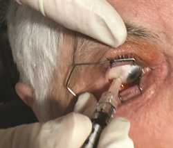
The eye and the eyelids are then cleaned, usually using povidone-iodine, a yellow solution which is very effective at killing bacteria that live around the eye. An eyelid speculum is often used to keep the eyelids open during the procedure. Once the eye is prepped for injection, you will be asked to look in a particular direction depending on the location of the injection while the medicine is injected through the pars plana (the white part of the eye) with a very small needle (Figure 1).
Typically, patients feel pressure, with little or no pain during the injection. After the injection, the speculum is removed and the eye is cleaned. The entire process takes about 10 to 15 minutes.
Safety and Results
Severe complications are very rare with intravitreal injections. The major risks are:
- Infection in the eye or endophthalmitis
- Inflammation in the eye or pseudoendophthalmitis (a non-infectious inflammatory reaction to some medications)
- Bleeding into the vitreous gel (vitreous hemorrhage)
- Retinal detachment
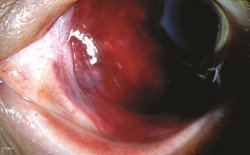
Sometimes there may be a small bleed or subconjunctival hemorrhage (Figure 2) on the surface of the eye where the needle enters; this usually heals within a week.
Your retina specialist may check the intraocular pressure (IOP), that is, the pressure within the eye, following the injection. There is a temporary rise in IOP that usually returns to baseline in a few minutes. The IOP may take longer to normalize in patients with glaucoma and needs to be monitored.
What to Expect After the Injection
There are usually no restrictions following the injection apart from avoiding potential contamination of the eye on the day of the injection. However, you should contact your retina specialist if you experience signs and symptoms of complications, such as:
- Eye pain or discomfort
- Increased floaters after the first day
- Increased sensitivity to light
- Decreased vision
Sometimes after an intravitreal injection, you may get the feeling that “something is in your eye”—this can be a reaction to povidone-iodine, which is used to clean the eye before the injection. Artificial tears (preferably sterile single use droppers) can be used to help ease symptoms of dryness and surface irritation.
A follow-up visit with your retina specialist will be scheduled depending on the disease being treated, but is usually about 4 to 6 weeks after the injection.
Repeated Intravitreal Injections
Intravitreal injections are an indispensable tool for the retina specialist to treat a variety of conditions. Their use has become much more common since the introduction of anti-VEGF medications in 2006. Intravitreal injections often need to be repeated in chronic conditions such as AMD, diabetic macular edema, and retinal vein occlusions, which require frequent office visits. Repeat injections are usually safely tolerated over several years. The need for a repeat injection is determined during the clinical examination, often with the use of diagnostic testing such as optical coherence tomography (OCT) and fluorescein angiography (FA).
Research is underway that will hopefully provide longer-acting treatments in the near future.
Authors
Thank You To The Retina Health Series Authors
Sophie J. Bakri, MD
Audina Berrocal, MD
Antonio Capone, Jr., MD
Netan Choudhry, MD, FRCS-C
Thomas Ciulla, MD, MBA
Pravin U. Dugel, MD
Geoffrey G. Emerson, MD, PhD
Roger A. Goldberg, MD, MBA
Darin R. Goldman, MD
Dilraj S. Grewal, MD
Larry Halperin, MD
Vincent S. Hau, MD, PhD
Suber S. Huang, MD, MBA
Mark S. Humayun, MD, PhD
Peter K. Kaiser, MD
M. Ali Khan, MD
Anat Loewenstein, MD
Mathew J. MacCumber, MD, PhD
Maya Maloney, MD
Hossein Nazari, MD
Oded Ohana, MD, MBA
George Parlitsis, MD
Jonathan L. Prenner, MD
Gilad Rabina, MD
Carl D. Regillo, MD, FACS
Andrew P. Schachat, MD
Michael Seider, MD
Eduardo Uchiyama, MD
Allen Z. Verne, MD
Yoshihiro Yonekawa, MD
Editor
John T. Thompson, MD
Medical Illustrator
Tim Hengst
Downloads
Copyright 2017 The Foundation of the American Society of Retina Specialists. All rights reserved.